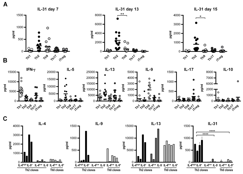
Figure 1. IL-31 secretion by in vitro differentiating CD4+ T-cell subsets and Bet v 1-specific T-cell clones.
Naïve CD4+ T cells were isolated from human buffy coats and cultured under Th1-, Th2-, Th9-, Th17-, and iTreg-polarizing conditions for 7 days, re-stimulated under the same conditions for an additional week and thereafter re-activated for 2 days with antibodies directed against CD3 and CD28. Supernatants were taken after each stimulation period and tested for IL-31 protein by ELISA. Data represent mean values of 13 polarization experiments carried out in duplicates using cells from 13 individual donors. (A). To control for the efficiency of CD4+ subset generation, signature cytokines for each subset were measured (B). T-cell clones specific for the major birch pollen allergen Bet v 1 and classified as Th2 and Th0 were stimulated with autologous irradiated PBMCs and 5μg/ml Bet v 1 for 48 hours. Secretion of the cytokines IL-4, IL-13 and IL-9 was determined using the Luminex System 100, and IL-31 expression was measured in duplicates by ELISA (C). Statistical analyses: one-way ANOVA/Dunnett’s multiple comparisons test. * p ≤ 0.05, ** p ≤ 0.01,**** p ≤ 0.0001.

