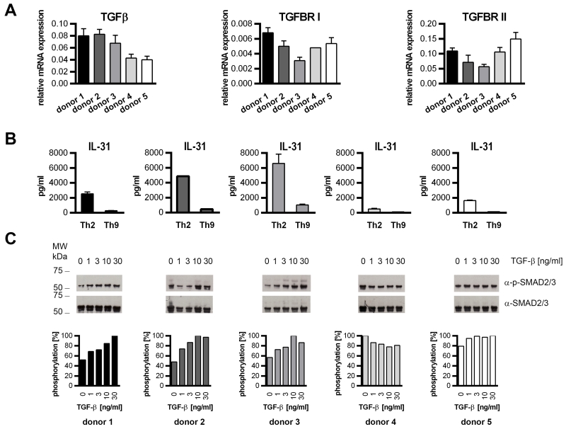
Figure 4. Phosphorylation of SMAD2 and SMAD3 correlates with limited IL-31 production.
A. Naïve human CD4+ T cells derived from 5 individual donors were analyzed for the expression of TGFβ, TGFBRI and TGFBRII mRNA by real-time q-RT-PCR. B. A fraction of the cells was in vitro-differentiated into Th2 and Th9 cells. IL-31 secretion was analyzed by ELISA on day 13. Data represent mean values of duplicates, error bars indicate standard deviations. C. Naïve CD4+ T cells were stimulated for 15 minutes with TGF-β1 at the concentrations indicated. Phosphorylation of SMAD2 and SMAD3 (p-SMAD2/3) and total SMAD2/3 was monitored by Western blotting. Phosphorylation levels relative to total SMAD2/3 and normalized to the signal maximum are shown.

