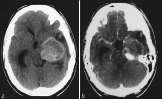
Figure 1.

(a and b) Cranial computed tomography in axial acquisitions showing an expansive lesion in the topography of the left posterior cerebral artery, with an important mass effect. In b, one can observe the partial opacification by means of a venous contrast
