Figure 3.

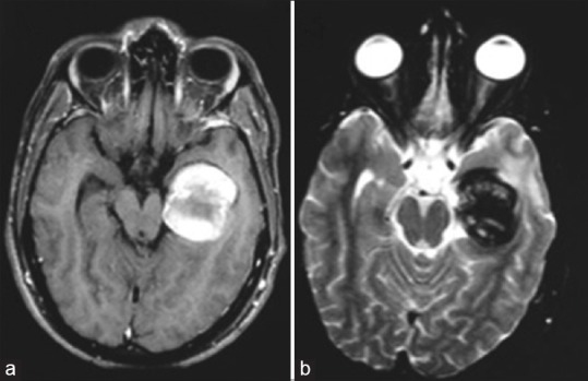
Cranial magnetic resonance imaging axial acquisition (a: T1-weighted; b: T2-weighted) showing an expansive lesion, with heterogeneous signal, suggesting a totally thrombosed giant aneurysm with occlusion of the posterior cerebral artery

Cranial magnetic resonance imaging axial acquisition (a: T1-weighted; b: T2-weighted) showing an expansive lesion, with heterogeneous signal, suggesting a totally thrombosed giant aneurysm with occlusion of the posterior cerebral artery