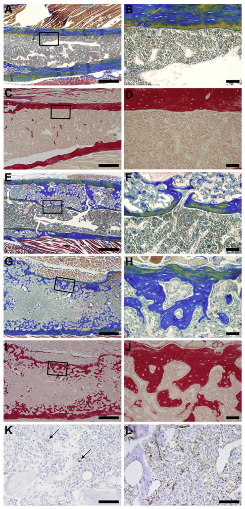
Fig. 2.
Femurs from an LP/J female (372 days, A–D, essentially normal bone and marrow), LP/J female (621 days, E, F, moderate, localized fibro-osseous lesions), and KK/HlJ female (586 days, G–J, severe diffuse fibro-osseous lesions) stained with Mallory’s (A, B, E–H) and Sirius Red (C, D, I, J). Note the decreasing thickness of cortical bone and increase in medullary trabeculae as the disease progresses. Left column 4× (bar=200 μm), right column 25× (bar=50 μm). Immunohistochemistry localization of CD31 for endothelial cells reveals few capillaries in the normal bone marrow of a 372 day old LP/J female compared to numerous vascular spaces in the 427 day old KK/HlJ female mouse with severe diffuse fibro-osseous lesions that completely efface the bone marrow cavity. 40×, Bar = 20 μm.

