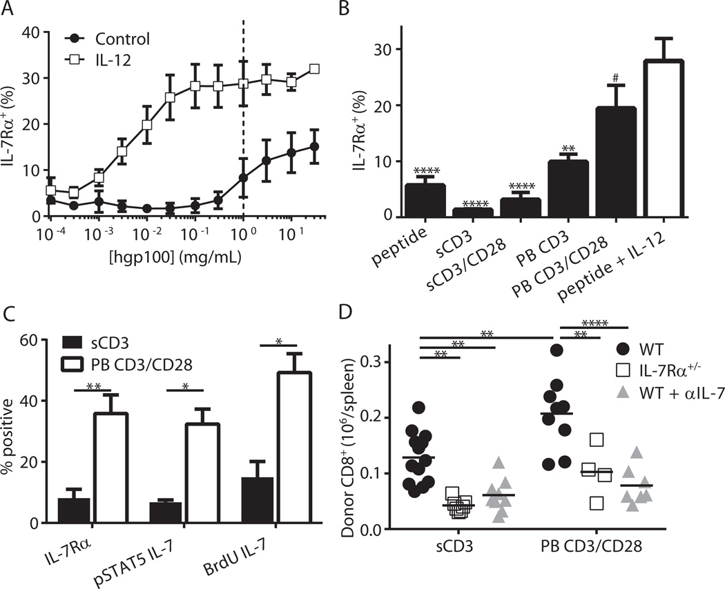
Figure 5. TCR strength modulates IL-7Rα expression, which dictates engraftment of activated CD8+ T cells.
(A) Pmel-1 CD8+ T cells were stimulated for 3 days ± IL-12 with titrated hgp100 peptide. (B) Pmel-1 T cells were stimulated with soluble anti-CD3 mAb (sCD3), sCD3 + soluble anti-CD28 mAb (sCD3/CD28), plate-bound anti-CD3 mAb (PB CD3), PB CD3 + plate-bound anti-CD28 mAb (PB CD3/CD28) or hgp100 peptide with or without IL-12 for 3 days and assessed for IL-7Rα expression (combined data from 4–5 independent experiments, # p > 0.05, ** p < 0.01, **** p < 0.0001 vs. hgp100 + IL-12). (C) B6 T cells were stimulated as indicated and assessed for IL-7Rα expression (n = 5, ** p < 0.01) or responsiveness to IL-7 (n = 3 for pSTAT5 and BrdU assays, * p < 0.05). (D) WT or IL-7Rα+/− mice were stimulated with soluble or plate bound antibodies then transferred into irradiated hosts. Where indicated, the IL-7 blocking antibody clone M25 was administered on days 0, 2 and 5 post-transfer. Shown are absolute numbers of donor CD8+ T cells 7 days after transfer (** p < 0.01, **** p < 0.0001, data is combined from 3 independent experiments).

