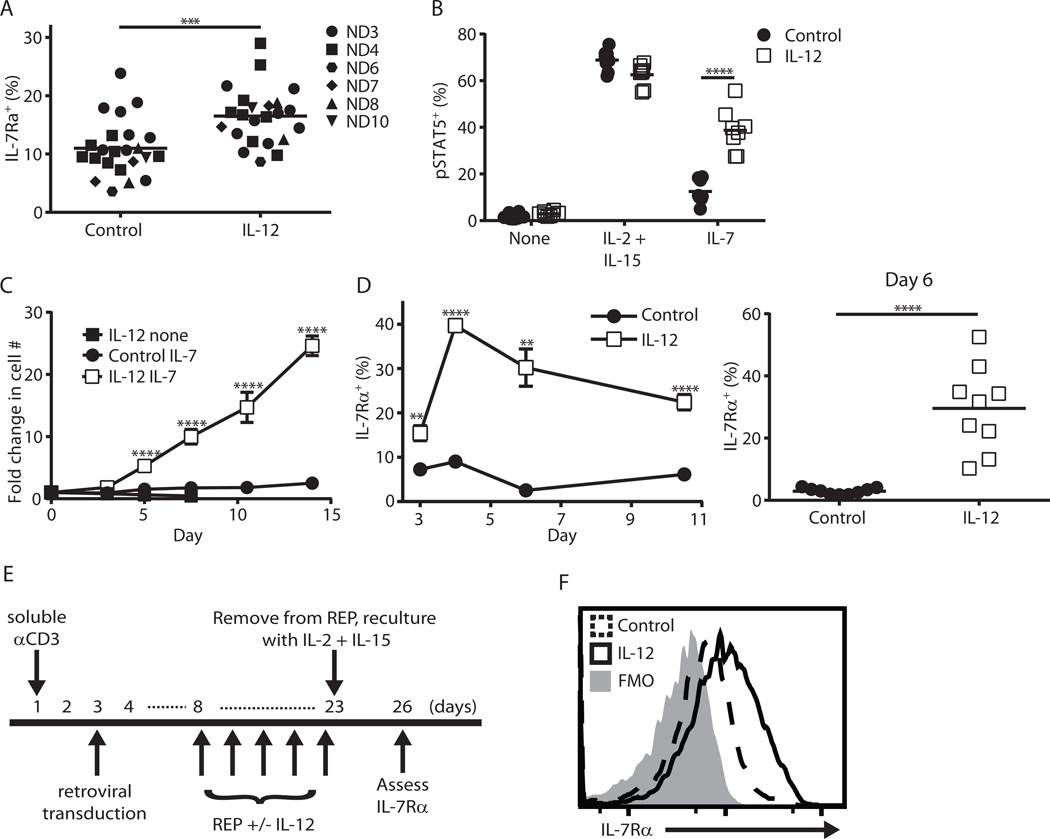
Figure 6. Human T cells conditioned with IL-12 display enhanced IL-7Rα expression and IL-7 responsiveness.
(A–D) Human PBMCs were activated with soluble anti-CD3 mAb (0.5 µg/mL, Okt3 clone) with or without hIL-12 (10ng/ml) for 3 days. (A) IL-7Rα expression after 3 day activation (*** p < 0.001; “ND” is normal donor). (B, C) Day 3 activated T cells were washed then replated in the indicated cytokines (300 IU/mL IL-2 + 100 ng/mL IL-15; IL-7, 100 ng/mL). (B) pSTAT5 staining via flow cytometry after overnight culture (n = 8 from 2 independent experiments with 4 normal donors, **** p < 0.0001). (C) Cells were counted and given fresh media every 2–3 days (n = 6 from 2 independent experiments with 3 normal donors). (D) As in (C) except activated cells were recultured in IL-2 + IL-15 on day 3 then assessed for IL-7Rα expression at the indicated time points (n = 6–9 from 2 independent experiments with 4 normal donors, ** p < 0.01, **** p < 0.0001 via Welch’s t-test). (E) Overview of the clinical transduction protocol to generate TCR-transduced melanoma-reactive human T cells. Shown is the timing of IL-12 addition and three day reculture in IL-2 (300 IU/mL) + IL-15 (100 ng/mL). (F) IL-7Rα expression at day 26 of above timeline of human T cells initially grown with or without hIL-12. This result is representative of two independent experiments.

