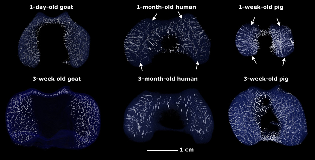
Fig. 1.

Three-dimensional reconstructions of susceptibility weighted magnetic resonance imaging data depicting the vascular architecture to the distal femoral epiphyseal cartilage in the axial plane in pigs (1 and 3 weeks old), human beings (1 and 3 months old) and goats (1 day and 3 weeks old). Note the presence of distinct axial (central) and abaxial (peripheral) vascular beds in humans and pigs with vessels oriented parallel to the articular surface creating a characteristic avascular region (white arrows) in the midsagittal plane in both condyles. In goats, the majority of the epiphyseal vessels arise from the ossification front, are shorter, and are oriented perpendicular to the articular surface. All specimens are oriented with the medial aspect towards the left and the anterior (cranial) aspect towards the top of the figure.
