Fig. 2.
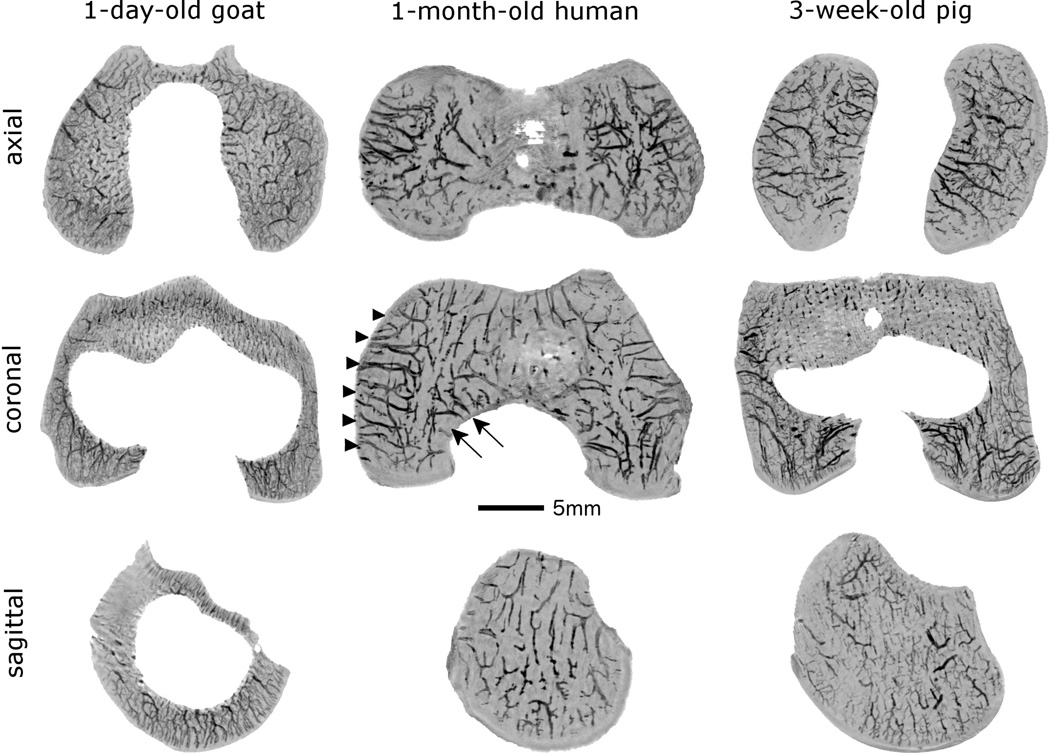
Four and five millimeter thick minimum intensity projections calculated from the susceptibility weighted imaging datasets in the coronal and axial planes demonstrate two distinct vascular beds (axial[central] and abaxial [peripheral]) in the epiphyseal cartilage of the femoral condyles in specimens obtained from a 1-month-old human and a 3-week-old pig (shown in the medial femoral condyle in the middle image, arrowheads mark abaxial, arrows mark axial vascular bed). Conversely, no separate vascular beds can be distinguished in the specimen obtained from a 1-day-old goat, where the majority of the vessels arise from the secondary ossification center providing a dense, evenly distributed vascular supply to both femoral condyles. In the two-millimeter-thick minimum intensity projection obtained in the sagittal plane the secondary ossification center is clearly apparent in the goat (white void in the middle of the image), consistent with more advanced skeletal development when compared to both the human and the porcine specimens. All specimens are oriented with the medial aspect towards the left of the figure. Red lines in the porcine specimen mark the plane for the images depicted for each species.

