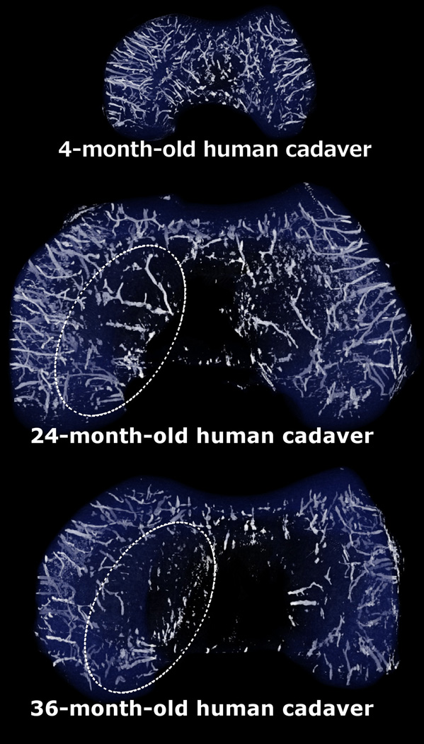
Fig. 3.
Three-dimensional reconstructions of the susceptibility weighted imaging data obtained from distal femoral specimens of 4-, 24- and 36-month-old human cadaveric donors demonstrating earlier resolution of the axial (central) vascular bed vs. the abaxial (peripheral) one. This area of decreased vascularity in the axial aspect of the femoral condyles (indicated with oval, dotted ellipses in the 24- and 36-month-old specimens) corresponds with the predilection site of osteochondrosis/osteochondritis dissecans. All specimens are oriented with the medial aspect towards the left of the figure.

