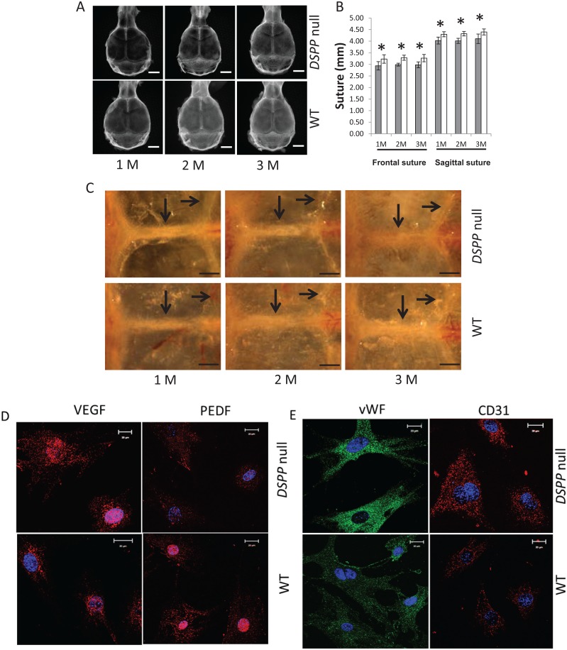
Figure 2.
Characterization of the calvarial bones in DSPP-null and gender-matched wild-type (WT) mice at 1, 2, and 3 mo by radiography and optical microscopy. (A) Representative radiographs reveal reduced mineralization of the calvarial bones. For radiographic imaging, the inferior side was facing the X-ray, and axial scans were obtained (n = 5). (B) Length of the frontal suture (FS) and sagittal suture (SS) from radiographic analysis. The mean ± standard deviation of measurements of the FS and SS from 5 samples for each group (*P < 0.05). Gray bar = DSPP null; white bar = WT. (C) Visualization through a dissection microscope of the coronal and sagittal sutures of the dissected skulls of 1-, 2-, and 3-mo-old WT and DSPP-null mice. Arrows point to the sutures. Note the increased vascularity of the parietal bones in the null mice. Bar = 1 mm. Arrows indicate frontal (long arrow) and lambdoid (short arrow) sutures. (D) Expression of angiogenic markers in the calvarial osteoblasts isolated from 7-d WT and DSPP-null mice. Anti-VEGF and anti-PEDF fluorescence immunocytochemistry performed on calvarial cells isolated from 7-d WT and DSPP-null mice. (E) Expression of endothelial markers in the calvarial osteoblasts isolated from 7-d WT and DSPP-null mice. Anti–von Willebrand factor and anti-CD31 fluorescence immunocytochemistry performed on calvarial cells isolated from 7-d WT and DSPP-null mice.

