Figure 1.
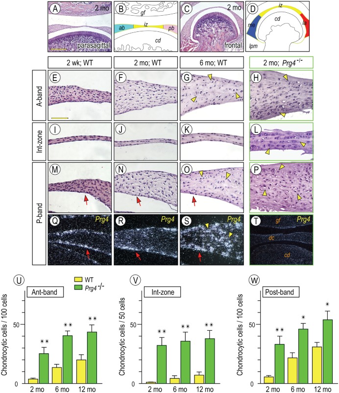
Temporospatial Prg4 expression in wild-type discs and aberrant differentiation of chondrocyte-like cells in Prg4-/- discs. Diagram (B, D) illustrating respective disc portions from parasagittal (A) and frontal sections (C) of temporomandibular joints (TMJs). TMJs from mice were analyzed by histology: 2-wk-old (E, I, M, Q), 2-mo-old (F, J, N, R), and 6-mo-old (G, K, O, S) control and 2-mo-old Prg4-/- (H, L, P, T). In situ hybridization with isotope-labeled riboprobes for Prg4 expression (Q–T). Note that Prg4 is expressed in disc-lining cells (arrows) and chondrocyte-like cells (arrowheads). Chondrocyte-like cells were counted in anterior (Ant) band, intermediate (Int) zone, and posterior (Post) band in distinct TMJ sections from control and Prg4-null mice at ages of 2, 6, and 12 mo (U–W; cell number is present per approximately 100 cells in anterior and posterior bands and approximately 50 cells in intermediate zone). Areas were randomly selected from 5 to 8 sections per sample (n = 3 for mouse/each group; *P < 0.05, **P < 0.01) and presented as average ± SD. Scale bars: 250 µm in A, also for C, T; 65 µm in E, also for F–S. ab, anterior band; cd, condyle; dc, disc; gf, glenoid fossa; iz, intermediate zone; la, lateral part; lpm, lateral pterygoid muscle; me, medial part; pb, posterior band; WT, wild type.

