Figure 2.
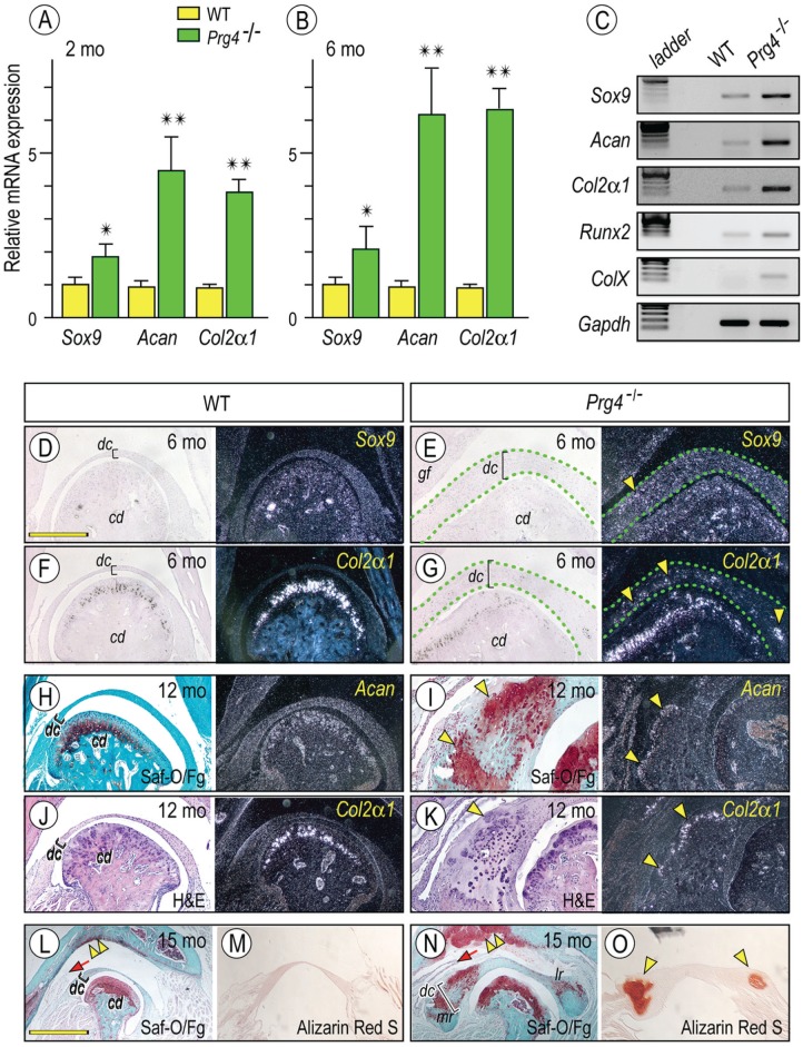
Increased expression of chondrocyte markers in Prg4-/- discs. Histograms depicting the relative expression of Sox9, aggrecan (Acan), and Col2α1 in Prg4-/- and control discs at 2 mo (A) and 6 mo (B) of age and presented as average ± SD (n = 3 for mouse/each group, *P < 0.05, **P < 0.01). Semiquantitative reverse transcription polymerase chain reaction reveals increased expression of early (Sox9, Acan, and Col2α1) and maturing chondrocyte (Runx2 and ColX ) markers in Prg4-/- discs as compared with control littermates at age of 6 mo (C). Frontal sections prepared from TMJs and discs from 6-mo-old (D–G), 12-mo-old (H–K), and 15-mo-old (L–O) control (D, F, H, J, L, M) and Prg4-/- (E, G, I, K, N, O) mice were processed for in situ hybridization with isotope-labeled riboprobes for Sox9 (D, E, arrowhead), Col2α1 (F, G, J, K, arrowhead), and Acan (H, I). Mutant discs were delineated by green dashed lines (E, G). Sections were also stained with safranin O (Saf-O) / fast green (Fg; H, I, L, N) and alizarin red S (M, O) and evaluate proteoglycans and/or mineralization in discs, respectively. Safranin O–stained cartilage (I, bright field, arrowheads) was composed of Col2α1- and Acan-expressing chondrocytes (I, K, dark field, arrowheads). Alizarin red S–stained mineralized tissues were detected in the mesial and lateral portions of the mutant discs (O, arrowheads). Note also the massive safranin O–stained cartilage developed in the mutant glenoid fossa as compared with control (L, N, double arrowhead, respectively) and the elongated medial synovial membrane in mutants (L, N, arrow). Scale bars: 1.2 mm in D, also for E–K, M, O; 2.5 mm in L, also for N. cd, condyle; dc, disc; gf, glenoid fossa; H&E, hematoxylin and eosin; WT, wild type.

