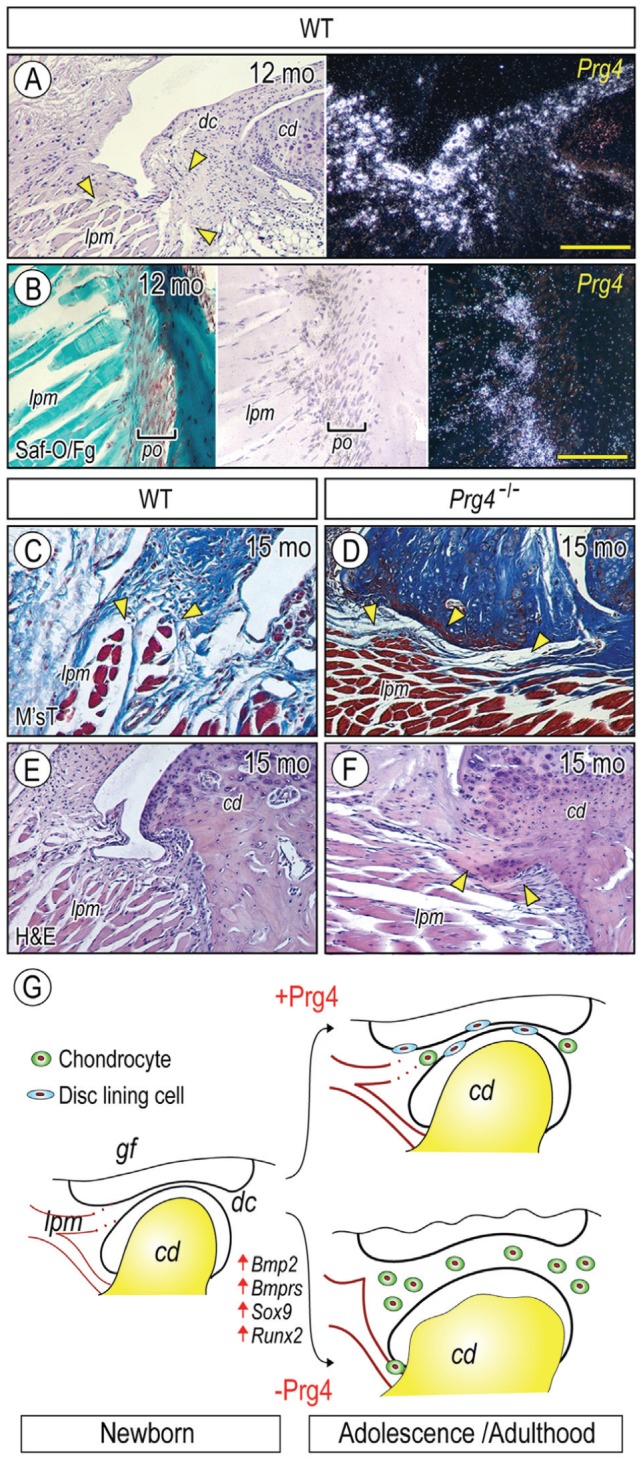
Figure 5.

Prg4 expression at nonarticulation sites and tissue derangement in Prg4-/- mice. Parasagittal sections of TMJs from 12-mo (A, B) and 15-mo (C–F) control (A, B, C, E) and Prg4-/- (D, F) mice were processed for histology and in situ hybridization with isotope-labeled riboprobes for Prg4 expression (A, B). Sections were stained with safranin O/fast green (Saf-O/Fg; B), Masson’s trichrome (M’sT; C, D), and hematoxylin and eosin (H&E; E, F). Note that Prg4 is expressed at disc/lateral pterygoid muscle junction (A, arrowheads) and muscle/tendon insertion site into the mandibular condyle (B). Note also the disrupted muscle insertion into the articular disc in mutant (D, arrowheads) as compared with control (C, arrowheads) and ectopic cartilage formation at muscle/tendon insertion site into the mandibular condyle in mutant (F, arrowheads). (G) Model of heterotopic ossification in TMJs caused by absence of Prg4 action and involvement of abnormal BMP signaling. (G) Model of increased chondrogenesis and subsequent cartilage formation in Prg4 mutant discs. TMJ appears to develop normally, and the disc separates the joint space into the upper and lower joint cavities. With further development, the disc displays a concaved shape; the disc-lining cells cover the surface of the disc; and a few chondrogenic cells and chondrocytes differentiate in the anterior and posterior bands in wild-type disc (with Prg4 action). In sharp contrast, in Prg4 mutant mice (without Prg4 action), the disc becomes much thicker, and this is accompanied by increased chondrocyte number and ectopic cartilage formation throughout the disc. In addition, the lateral pterigoid muscle loses its smooth integration into the disc. The increased chondrogenesis and subsequent cartilage formation throughout the disc are accompanied by activation of Bmp signaling and increased expression of Sox9 and Runx2. Scale bars: 120 µm in A, also for E, F; 60 µm in B, also for C, D. cd, condyle; dc, disc; lpm, lateral pterygoid muscle; po, periosteum.
