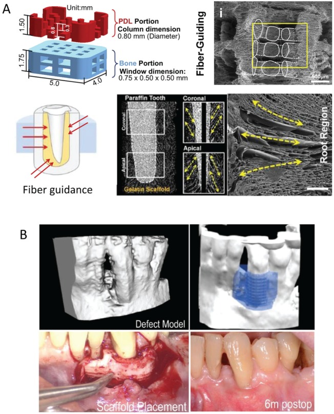
Figure 4.
Design of a customized scaffold using 3-dimensional (3D) printing. (A) 3D printing has been used to design a scaffold consisting of a periodontal ligament (PDL) portion and a bone portion. This was further modified to improve the fiber-guiding potential as well as the direction of the PDL to mimic the topography of the different kinds of fibers in the PDL (first row). Digitalized cross-sectional view of a 3D-reconstructed image. Longitudinal cross-section image showed pore morphologies at coronal and apical portions. Scanning electron microscopy image providing longitudinal pores produced by a freeze-casting method. Adapted from Park et al. (2010), Park et al. (2012), and Park et al. (2014) with permission. (B) 3D printing using polycaprolactone was made to fit the periosseous defect based on the patient’s cone beam computed tomography scan. Adapted from Rasperini et al. (2015) with permission.

