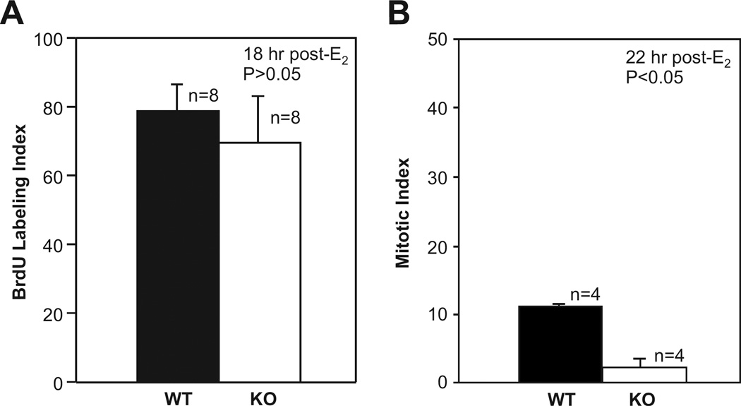
Fig. 2. BrdU uptake and mitosis in uterine epithelial cells of estradiol-treated wild-type and irs1−/− mice.
(A) Mice were treated with estradiol and euthanized 18 h later. BrdU was injected two hours prior to euthanasia. Standard methods were used to prepare uterine sections for examination by light microscopy. BrdU was detected in epithelial cells with a rat anti-BrdU monoclonal antibody.
(B) Mitosis in uterine epithelial cells was determined by injecting demecolcine into mice 20 h after hormone treatment, followed by euthanasia at 22 h. Further details of the procedures used are described in Materials and Methods. In A and B, each bar represents the mean ± standard deviation; n is the number of mice used in each group. For both BrdU uptake (A) and mitosis (B), approximately 500 epithelial cells were counted in each uterine section. The mitotic index of IRS-1-knockout (KO), or irs1−/− mice, was significantly different from that of wild-type at p < 0.05.

