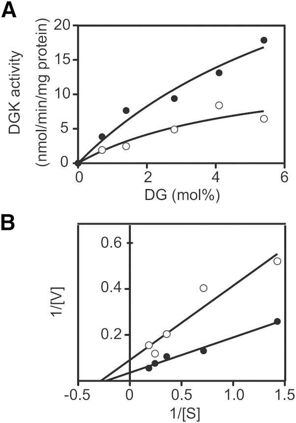
Fig. 6.
Effect of CU-3 on the affinity of DGKα for DG. A: The 12,000 g supernatant (5 μg) of the extracts from COS-7 cells expressing DGKα was incubated with various concentrations of DG as indicated for 5 min in the presence (1 μM, open circle) or absence (DMSO alone, closed circle) of CU-3. B: Lineweaver-Burk plots of the data. The values are the averages of duplicate determinations. The data shown are representative of three separate experiments.

