Fig. 8.
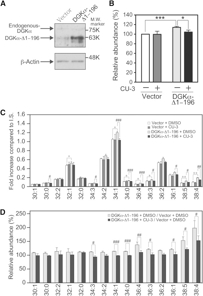
Effect of CU-3 on the DGKα activity in cells. COS-7 cells were transfected with p3xFLAG-DGKα-Δ1–196 or p3xFLAG vector alone (Vector). After a 24 h incubation, 2 μM of CU-3 or DMSO alone was added and further incubated for 24 h. A: Expression of endogenous DGKα and DGKα-Δ1–196 in COS-7 cells. COS-7 cells transfected with p3xFLAG-CMV vector alone or p3xFLAG-CMV-DGKα-Δ1–196 were harvested, and the cell lysates (15 μg of protein) were analyzed by Western blotting using anti-DGKα antibody. B–D: The amounts of the total PAs (B) and major PA molecular species (C) in COS-7 cells were quantified using the LC/MS method. The values are presented as the mean ± SD (n = 3). B: * P < 0.05, *** P < 0.005. C: * P < 0.05, ** P < 0.01 (p3xFLAG-vector-transfected cells in the absence of CU-3 vs. p3xFLAG-DGKα-Δ1–196-transfected cells in the absence of CU-3); # P < 0.05, ## P < 0.01, ### P < 0.005 (p3xFLAG-DGKα-Δ1–196-transfected cells in the absence of CU-3 vs. p3xFLAG-DGKα-Δ1–196-transfected cells in the presence of CU-3). p3xFLAG-vector-transfected cells in the absence of CU-3, white bars; p3xFLAG-vector-transfected cells in the presence of CU-3, dark gray bars; p3xFLAG-DGKα-Δ1–196-transfected cells in the absence of CU-3, light gray bars; p3xFLAG-DGKα-Δ1–196-transfected cells in the presence of CU-3, black bars. (D) The results are presented as the relative value of major PA molecular species of p3xFLAG-DGKα-Δ1–196-transfected cells versus p3xFLAG vector-transfected cells in the presence (black bars) or absence (white bars) of CU-3. # P < 0.05, ## P < 0.01, ### P < 0.005.

