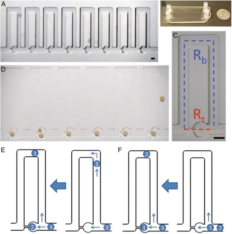
Figure 3.
The simple perfusion apparatus (SPA) using a hydrodynamic trap array: (A) images of the 8-trap hydrodynamic array. Oocytes flow in and load into the wells from right to left, which is the direction of fluid flow. (B) Macroscale view of hydrodynamic trap arrays. Image displays two parallel arrays bonded to a standard size microscope slide coverslip. (C) An individual trap. This consists of a trapping channel (red) and a bypass channel (blue). The resistances of each segment dictate the trapping behavior of the device. The trapping region consists of a circular trap (320 μm in diameter) and a high resistance channel (40 μm width), which connects to the ends of the bypass segment. (D) Immature sea star oocytes within the chip. Oocytes load into the chip from right to left for the six traps displayed. The nucleus and nucleolus are visible in each oocyte which is ∼200 μm. Double trapping can occur with smaller oocytes, as seen in trap five. Scale bars = 200 μm. (E) Indirect trapping. Occurs when the trap resistance is greater than the bypass channel resistance (Rt:Rb = 1.56) causing the first oocyte to be directed into the bypass channel. (F) Direct trapping. Occurs when the trap resistance is lower than the bypass resistance (Rt:Rb = 0.8), directing the first oocyte into the trap. Subsequent oocytes can be directed into the trap causing double trapping, or directed into the bypass channel depending on the change in resistance due to the trapped oocytes for either method (from Angione et al., 2015).

