
Extended Data Figure 7. PVT1 regulates MYC protein level in MDA-MB-231 breast cancer cells.
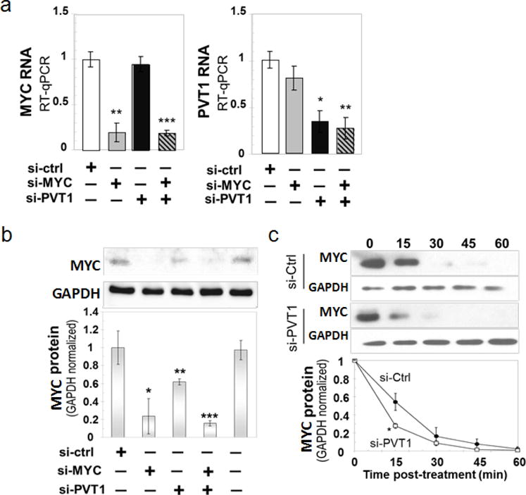
a, RT–qPCR measurement of MYC (left) and PVT1 (right) RNA levels in MDA-MB-231 cells 48 h after transfection with the indicated siRNAs. b, Reduction in MYC protein in PVT1-depleted MDA-MB-231. Western blot analysis for MYC protein in the total lysates obtained from the MDA-MB-231 cell line transfected with different siRNAs. The relative density for each category was determined by normalizing against the intensity of the GAPDH band. c, Stability assay for MYC protein in MDA-MB-231 cells. Cells were transfected with siCtrl and siPVT1 and then treated with 10 μM cycloheximide for different time points (top panel). The relative density was determined by comparing against the GAPDH level (bottom panel). Mean ± s.e.m. for a–c (n = 3). *P < 0.05, **P < 0.01, ***P < 0.001 by two-tailed Student’s t-test. Error bars, s.e.m.