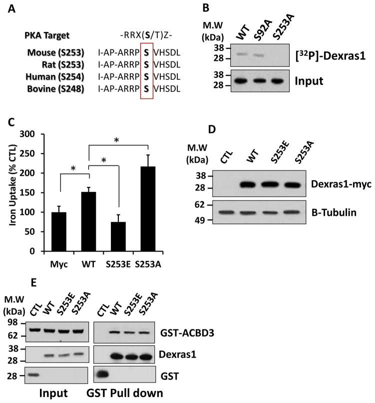
Figure 2. Serine 253 in Dexras1 is phosphorylated by PKA.
(A) Amino acid sequence alignment of Dexras1. (B) Either WT Dexras1-myc or phosphodead mutants (S92A or S253A) were transfected into HEK293T cells and were immunoprecipitated by anti-myc antibody. In vitro phosphorylation assay was performed and the results were visualized by autoradiography (top). Western blot image shows the loading of Myc-tagged Dexras1 protein in the reactions (bottom). (C) HEK293T cells were transfected with WT, S253E or S253A mutant Dexras1 and were subjected to iron uptake assays. (D) HEK293T cells transfected with WT, S253E or S253A mutant Dexras1 were subjected to western blotting. (E) HEK293T cells were transfected with GST-tagged Dexras1 (WT, 253E or S253A) and ACBD3. GST-pull down assay was performed. Immunoassays were performed three times. Iron uptake experiments were repeated five times, each sample in at least triplicate.

