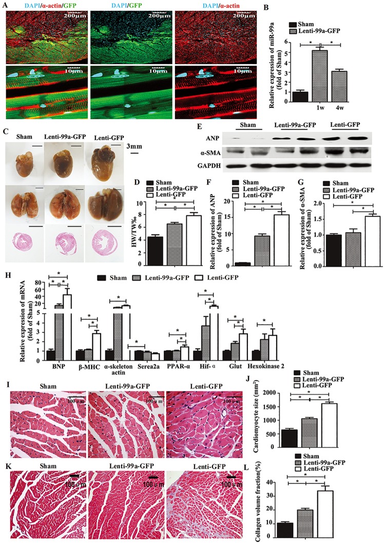
Fig 4. MiR-99a gene therapy attenuates cardiac hypertrophy in a mice model with TAC.
A, Immunofluorescence for GFP tag, indicating successful expression of exogenous miR-99a in heart after lenti-99a-GFP delivery. (α-actin: red; GFP: green; DAPI: cyan). B. Increased miR-99a expression in heart 1 week and 7 weeks after lentiviral delivery assessed by TaqMan RT-PCR. C More spherical shape of hearts from lenti-GFP mice (right column) comparing to lenti-99a-GFP (middle column) and sham mice (left column). Bigger size of lungs from lenti-GFP mice (right column) relative to lenti-99a-GFP (middle column) and sham mice (left column). The bottom panel shows paraffin-embedded sections of the hearts from the top panel. Bars = 3mm. D. Heart-to-body-weight ratio (mg/g) was increased in TAC mice, but attenuated in miR-99a overexpressing heart (n = 8 in lenti-99a-GFP group; n = 19 in lenti-GFP group). E-G. Western blotting analysis showed ANP (F) and α-SMA (G) were both down-regulated in miR-99a treated group compared to lenti-GFP group. H. BNP, β-MHC, PPAR-α, ACTA1, Hif-α, GLUT1 and HK2 levels were strongly increased in lenti-GFP treated hearts, but attenuated in lenti-99a treated hearts. Serca2a was decreased after surgery, but there was no significant difference in Serca2a expression among these three groups. I-J. Analysis of cardiomyocytes size in HE—stained sections (n = 3–5 per each group). K-L. Analysis of cardiac fibrosis in Masson—stained sections (n = 3–5 per each group). *, p<0.05.

