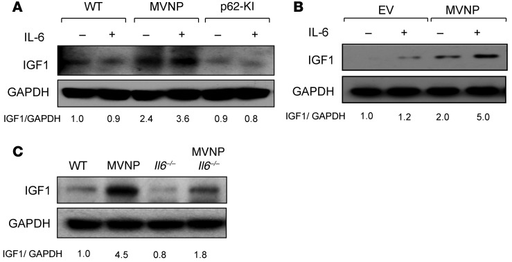
Figure 6. IL-6 increases IGF1 expression in MVNP-expressing OCLs.
(A) OCLs formed by CD11b+ cells from 8-month-old WT, MVNP, and p62-KI mice were treated with IL-6 (10 ng/ml) for 4 days and cell lysates analyzed for IGF1 expression. (B) OCLs from normal human OCL precursors transduced with EV or MVNP retroviral constructs were treated with IL-6 (10 ng/ml) for 4 days and assayed for IGF1 expression. Results shown in B are derived from the same gel shown in Figure 4E. (C) IGF1 expression by OCLs formed by CD11b+ cells from 8-month-old mice. The basal ratio of every molecule/loading control for vehicle treatment of WT cells was set at 1.0 in A–C. Results are representative of 3 biological replicates.

