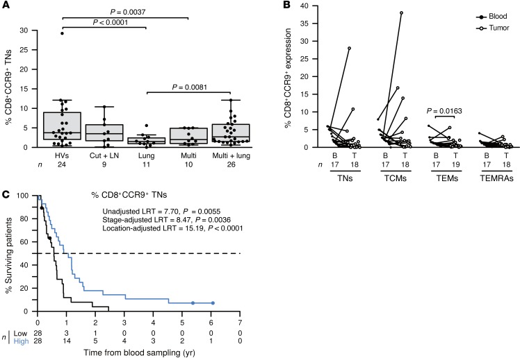
Figure 4. CD8+CCR9+ TNs leave the blood during lung metastasis and dictate MMel prognosis.
(A) CCR9 expression on CD8+ TNs is depicted for HVs, patients presenting with only cutaneous/LN (Cut + LN) metastases, those with additional lung (Lung) involvement, those with disseminated disease (Multi), and those with distant metastases plus lung involvement (Multi + lung) at the time of inclusion. Box plots summarize data from 57 patients with MMel and 24 HVs. (B) Match-paired comparison of CCR9 expression levels (performed by flow cytometry on fresh tissues) in all CD8+ T cell subsets from blood and tumors at the time of surgery in the prospective cohort of 20 patients with MMel. (C) Kaplan-Meier survival curves of 57 patients with MMel according to their proportions of circulating CCR9+CD8+ TNs segregated with the median. Each point represents 1 patient specimen, and the total number is indicated for all subpopulations studied. Statistical analyses were performed by beta regression (A), linear mixed-effects (B), and Cox regression (C) modeling. Raw P values are indicated.

