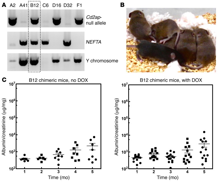
Figure 2. Development of ES cells sensitized for FSGS.
(A) Identification of FSGS-sensitized ES cells. Our breeding strategy predicted that 1 of 8 embryos would have the correct genotype. ES cells were generated using standard approaches and genotyped for Cd2ap heterozygosity (upper panel), the NEFTA transgene (middle panel), and the Y chromosome (lower panel). (B) Laser-assisted injection generated mice with high chimerism. In the example shown, the ES cell line (agouti) was injected into 8-cell C57/BL6 (black) embryos. Compared with noninjected embryos (resulting in the 2 black mice shown on the bottom), all of the injected embryos generated pups that were close to being purely agouti. Injection of ES cells into C57/BL6 albino embryos resulted in completely agouti animals (not shown). (C) Mice generated from ES cells developed mild proteinuria after 4 months, with no DOX treatment. Fifteen mice were generated from the sensitized ES cells and treated with or without DOX in the drinking water. Urine was tested every month by measuring the albumin/creatinine ratios. Mice developed low-level proteinuria at 4 months of age, but the level of proteinuria was not affected by DOX treatment.

