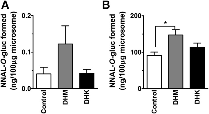
Fig. 7.
NNAL-O-gluc formation by (A) lung microsomes and (B) liver microsomes from A/J mice exposed to dietary DHM or DHK in comparison with control (n = 5). We incubated 5 mM NNAL for 30 minutes with NNAL-O-gluc quantified by LC-MS/MS. Statistical analysis was performed with one-way analysis of variance followed by Dunnett’s test relative to the control group; *P < 0.05.

