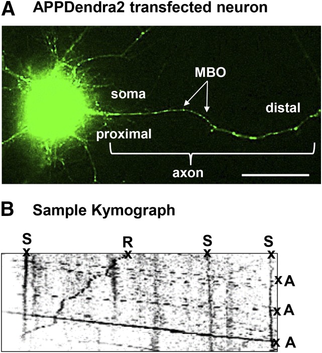
Fig. 1.
Methods for assessing the effects of diisopropylfluorophosphate (DFP) on axonal transport in vitro. (A) Representative image demonstrating successful transfection of APPDendra2 in rat primary cortical neurons. Green fluorescent MBOs in the axon are indicated by the arrows. (B) Kymograph recorded at 5-second intervals for 5 minutes generated from APPDendra2-labeled MBOs after treatment with DFP. MBOs are categorized in one of three ways: anterograde (A), retrograde (R), or stationary (S). Velocity information was obtained from the slope of the lines. Scale bar = 20 µm.

