Fig. 3.
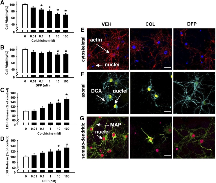
Twenty-four hours of exposure to colchicine (COL) and diisopropylfluorophosphate (DFP) are associated with concentration-dependent impairments in cell viability. After 24 hours of exposure to COL (A, C) or DFP (B, D), cell viability was measured by both MTT colorimetric and LDH release assays, presented as the percentage of cell survival or release, respectively, compared with vehicle-treated controls. Each bar represents the mean ± S.E.M. (n = 2 to 3 independent experiments). *P < 0.05 compared vehicle control conditions. Representative immunocytochemistry images (pseudo-colored) for contrast and to facilitate identifying stains in the figure and indicated structures) comparing the cellular morphology and structure after 24 hours of exposure to 10 μM COL or DFP. (E) Fluorophore-conjugated actin (Acti-stain 555, red) and nuclei (blue). (F) Antidoublecortin (DCX, axon, blue) and nuclei (yellow). (G) Antimicrotubule-associated protein 2 (MAP2, somatodendritic, green) and nuclei (red). Scale bar = 10 µm.

