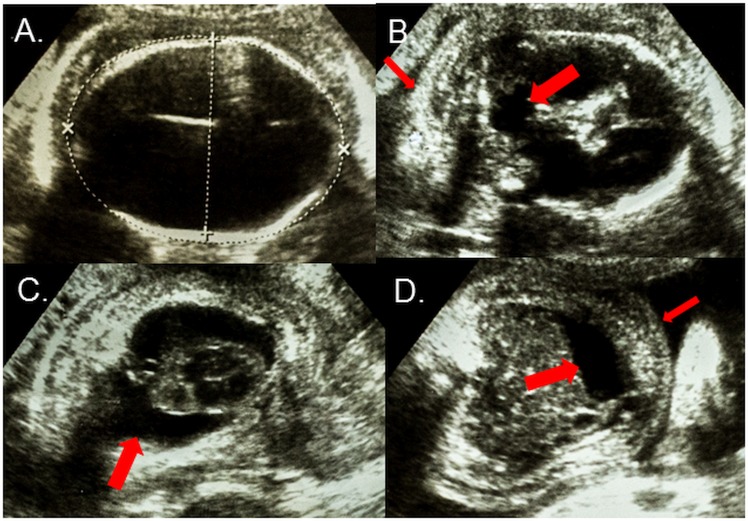
Fig 1.
Axial ultrasound views of the fetus at the 30th gestational week showing (A) Cranium with severe microcephaly (215mm) and hydranencephaly; (B) Posterior fossa with destruction of the cerebellar vermis (wide arrow) and nuchal edema (thin arrow); (C) Thorax with bilateral pleural effusions (arrow); and (D) Abdomen with ascites (wide arrow) and subcutaneous edema (thin arrow).

