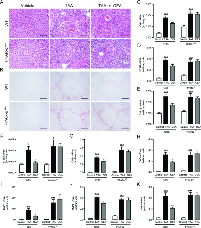
Figure 4. Anti-fibrotic effects of OEA in TAA-induced fibrosis mice were mediated by PPAR-α activation.
A.-B. Hematoxylin-eosin (HE) and Sirius red staining of liver sections. C.-K. Hepatic mRNA levels of ICAM, VCAM, TGF-β, α-SMA, Col1a, Col3a, TIMP1, MMP-2, and MMP-9 were determined by quantitative real-time PCR analysis. Results from wild-type (WT) mice and PPAR-α knockout mice with saline treatment, TAA treatment (160 mg/kg, i.p.), TAA treatment combined with OEA administration (5 mg/kg/day, i.p.). Data are shown as means ± s.e.m.; n = 6-8 in each group. ## P < 0.01, ### P < 0.001, * P < 0.05, ** P < 0.01, *** P < 0.001. Scale bars: 100 μm (A), 200μm (B).

