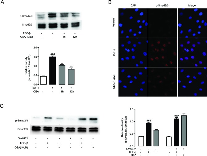
Figure 6. Inhibitory effect of OEA on TGF-β1-dependent Smad2/3 phosphorylation in vitro was mediated by PPAR-α.
A. CFSC cells were treated with TGF-β1 (5 ng/mL) for 30 min after 1 h or 12 h OEA (10 μM) treatment, Smad2/3 phosphorylation was analyzed by Western Blot. B. CFSC cells were treated with TGF-β1 (5 ng/mL) for 30 min after 1 h OEA (10 μM) treatment, phosphorylation of Smad2/3 was analyzed by Immunofluorescence staining. C. CFSC cells were treated with TGF-β1 (5 ng/mL) for 30 min after 1 h OEA (10 μM) treatment in the absence or presence of GW6471 (10 μM). Data are shown as means ± s.e.m of three independent experiments. ### P < 0.001, ** P < 0.01, *** P < 0.001.

