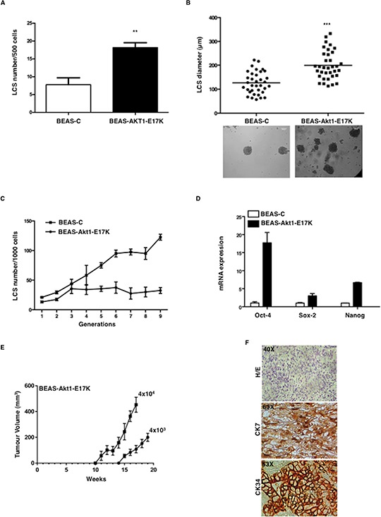
Figure 1. Mutant Akt1-E17K increases formation of LCSs.

A. Number of primary LCSs generated from control BEAS-2B cells or from the corresponding cells infected with pLenty-Akt1-E17K. **p < 0.01. B. Analysis of size distribution (μm) of LCSs generated from control BEAS-C and BEAS-Akt1-E17K cells by phase-contrast microscopy ***p < 0.001. C. Number of LCSs generated from control BEAS-C and BEAS-Akt1-E17K cells during serial passages expressed as mean ± SD. D. Relative mRNA expression of stemness genes by Q-RT-PCR in BEAS-C and BEAS-Akt1-E17K cells. E. Tumor growth of primary LCS generated from BEAS-Akt1-E17K cells (4 × 10³, 4 × 10⁴), injected into the flank of NOD/SCID mice (n = 8/group); data are shown as mean ± SD. F. Representative images of CK7 and CK34 immunostaining of tumours generated from single cell suspensions of primary LCSs derived from BEAS-Akt1-E17K cells subcutaneously injected into the flank of NOD/SCID and explanted 15–20 weeks after injection. Magnification as indicated.
