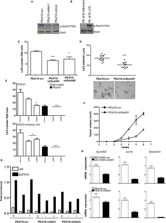
Figure 7. LCS formation and tumorigenicity of NSCLC cells are regulated through STAT3 activity.

A. Immunoblot analysis of phosphorilated and total STAT3 (pY705) in PEd/10-scr, PEd/10-shAkt1 and PEd/10-shIL-6r cells. B. Immunoblot analysis of phosphorylated (pY705) and total STAT3 in PEd/10-scr cells grown as LCS or in adherent conditions. C. Number of primary LCSs generated from PEd/10 cells or the corresponding cells interfered for STAT3 (PEd/10-shSTAT3; clones #1, #2). ***p < 0.001, *p < 0.05. D. Analysis of size distribution (μm) of LCSs generated from PEd/10 cells and derivatives by phase-contrast microscopy ***p < 0.001. E. Analysis of spheroid forming capability of NCI-H460 cells and PEd/10 cells in the presence of Sta-21 and Stat3 Inhibitor XIII at the indicated concentrations (upper and lower panels, respectively). ***p < 0.001, *p < 0.05. F. Tumor growth of PEd/10-scr and the corresponding cells interfered for STAT3 injected into immunodeficient mice (n = 5/group). G. STAT3 occupancy (fold enrichment) of JunB promoter as determined by Chip of cross-linked DNA extracted from control PEd/10-scr, PEd/10-shAkt1 and PEd/10-shSTAT3 cells growing in complete (C), serum free (−) and serum free plus IL-6 (+) medium using anti-STAT3 or control IgG isotype antibodies. H. Relative mRNA expression of STAT3 target genes in PEd/10-scr and PEd/10-shAkt1 cells by Q-RT-PCR analysis.
