
Figure 4. Effects of 2CP on tail bleeding time and mouse pulmonary metastasis.
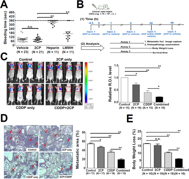
A. The mice were intravenously injected with the control vehicle DMSO, 2CP (3.5 mg/kg), heparin (2 mg/kg) or LMWH (2 mg/kg) followed by measurement of the mouse tail bleeding time. The bleeding time was plotted and expressed as the mean ± S.E. A bleeding time longer than 360 sec was set as 360 sec. B. Timeline for the experimental protocols of the mouse pulmonary metastases model. The administration schedule and the therapeutic efficacy for 2CP and CDDP (2.5 mg/kg) were analyzed at the indicated time points. C–E. Representative bioluminescence images for tumor growth were shown (panel C, left) and quantified (panel C, right). Representative images of lung sections with metastatic foci (Hematoxylin and Eosin stain) were analyzed and quantified using Zeiss Axiovision software. Arrows point out the metastatic foci. (100 X magnification, scale bar = 100 μm). The corresponding metastatic area was expressed as the percentage of the whole lung region (panel D). The body weight was recorded and the percentage of body weight loss at day 21 after tumor inoculation was calculated. Data represent the mean ± S.E. from three to five independent experiments (panel E). *P < 0.05 and **P < 0.01 when compared with the control treatment. n.s., no significance.