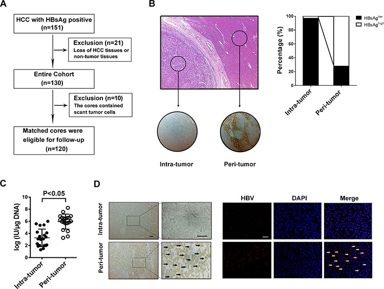
Figure 1. HBV infection in HCC tissues is lower than that in the surrounding liver tissue.

A. Patient demographic and baseline characteristics. B. Tissue microarray section contains tumor tissue and paired-surrounding tissue identified by H&E staining, and immunostaining with anti-HBsAg antibody shows that HBsAg expression was mainly localized to the cytoplasm and membrane (right); Of the 120 pairs samples, the percentage of HBsAg-high staining in intra-tumor is less than in peri-tumor (left). C. RT-PCR is used to detecte HBV DNA load fresh HCC tissue and matched surrounding tissue, and the results show that the median DNA level is much lower in tumor tissue than in peri-tumor tissue (P < 0.001). D. The specific whole length HBV DNA is used as probe to perform FISH on paraffin tissue samples, and the surrounding normal liver cells present positive signals for HBV DNA, but most tumor cells don't have FISH signal no matter with Diaminobenzidine (left) or immunofluorescence staining (right).
