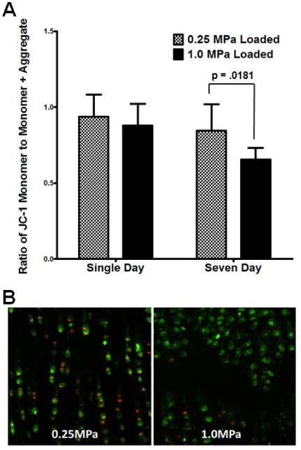
Figure 4. Mitochondrial membrane potential is depressed after one week of injurious loading.

Sagittal sections of the loaded area of each explant were assayed immediately after harvest for mitochondrial membrane potential using JC-1 confocal microscopy. Similar to respiratory effects, single day overloading has no impact upon mitochondrial membrane potential compared to normally loaded controls (quantified in A, representative pictures in B). By contrast, overloading for one week appears to depress mitochondrial membrane potential to levels consistent with the losses in mitochondrial function described above (18). Data represent the mean of at least three images per sample, standard error of the mean of n = 4 is shown, p-values as indicated. These data suggest that the losses in respiration observed in extracellular flux measurements are present immediately at harvest and also indicate a disruption of overall mitochondrial function consistent with osteoarthritis.
