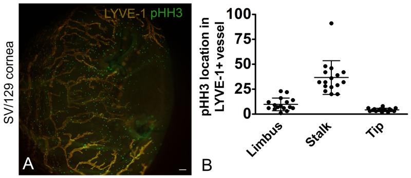
Figure 7.
Proliferating LEC stalk cells. Lymphangiogenesis was stimulated in SV-129 mice using the suture induced model of corneal inflammation. Corneas were labeled with antibodies to LYVE-1 and pHH3. An image obtained using epifluorescent microscopy is shown (A). Single z plane images were obtained using confocal microscopy from 14 individual mice. Random fields containing LYVE-1+ lymphatic vessels were identified and the localization of the pHH3 staining (limbus, stalk, tip) was quantified. pHH3 staining was the greatest in the LEC stalk cells (B). The data shown in B is composite data from 3 independent studies with 6 mice in each group, dots represents events per field. The size standard is 100 μM.

