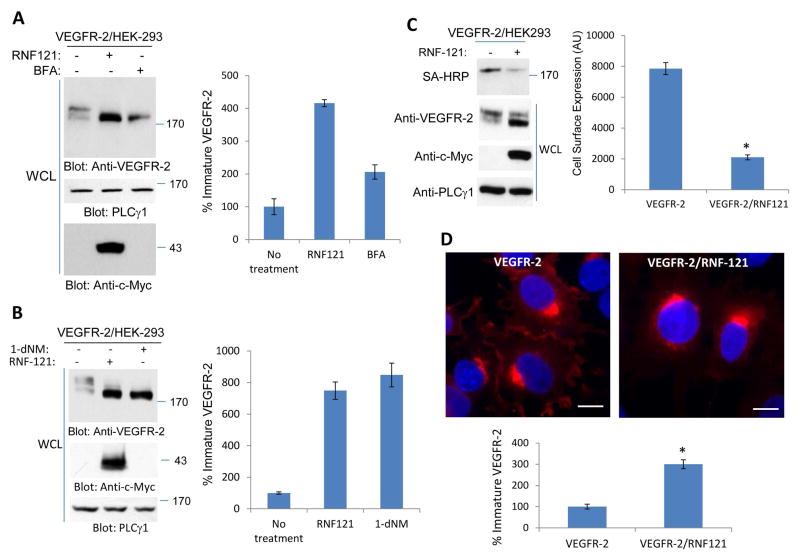
Figure 2. RNF121 regulates the cell surface levels of VEGFR-2.
(A) HEK-293 cells expressing VEGFR-2 alone or VEGFR-2 with c-Myc-RNF121 were treated with Brefeldin-A (BFA) or control vehicle and cells were lysed and whole cell lysates (WCL) were blotted for VEGFR-2, RNF121 and PLCγ1 as a loading control. (B) The same cell lines were treated with 1-deoxynojirimycin (1-dNM) and whole cell lysates were blotted for VEGFR-2, RNF121 and PLCγ1 as a loading control. (C) HEK-293 cells expressing VEGFR-2 alone or VEGFR-2 with c-Myc-RNF121 were subjected to cell surface biotinylation assay and biotinylated proteins was detected with Streptavidin-HRP (SA-HRP) as described in the Materials and Method section. Whole cell lysates were also blotted for VEGFR-2, RNF121 and PLCγ1. Shown is the quantification of the blot of cell surface biotinylation of VEGFR-2. *P<0.035. n=3 (D) Immunofluorescence staining of HEK-293 cells expressing VEGFR-2 or VEGFR-2 with c-Myc-RNF121 is shown. Cells were stained with anti-VEGFR-2 antibody. Scale bars, 10μm. (E) Graph is representative of immature VEGFR-2 from quantification of at least 20 cells per group. The Image J software was used to quantify the images. P<.005, n=3.

