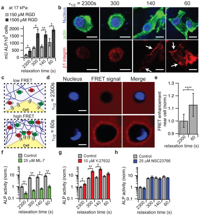
Figure 4. Osteogenic differentiation of MSCs mediated through ECM ligand density, enhanced RGD ligand clustering, and myosin contractility in stiffer hydrogels.
a, Quantification of ALP activity of MSCs encapsulated in hydrogels with an initial elastic modulus of 17 kPa after 7 days in culture with an RGD density of 150 or 1500 μM. b, Representative immunofluorescence staining for actin (green), nucleus (blue), and β1 integrin (red) in MSCs cultured in the indicated conditions for a week. c, schematic of assay using FRET between RGD-fluorescein and RGD-rhodamine coupled to different alginate chains to monitor mechanical clustering of RGD ligands at the nanoscale by cells locally. d, Representative confocal microscope images of nucleus (DAPI/ blue) and FRET acceptor signal from hydrogel (red) surrounding MSCs cultured in hydrogels with different stress relaxation properties after 18 hours of culture. Blank spot in FRET signal images indicates location of cell. e, Quantification of enhancement of FRET acceptor signal within ~2 – 3 um of cell border relative to the background of the hydrogel. Data are shown as mean +/− s.d. and **** indicates p < 0.0001 (student’s t-test). f, ALP activity of MSCs in the presence of ML-7, a myosin light chain kinase inhibitor. g, ALP activity of MSCs in the presence of a Rho kinase inhibitor, Y-27632. h, ALP activity of MSCs in the presence of a Rac1 inhibitor, NSC 23766. All experiments were done in hydrogels with an initial elastic modulus of 17 kPa and RGD concentration of 1500 μM. All data are shown as mean +/− s.d. *,** indicate p < 0.05, 0.01 respectively (student’s t-test). Scale bars are all 10 μm.

