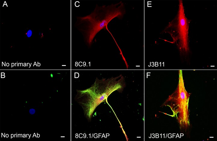
Fig 6. CR1 expression by human AD brain-derived astrocytes.
Representative confocal photomicrographs of astrocytes isolated from post-mortem human brain stained with 8C9.1 (red) (C, D) and J3B11 (red) (E, F) mAbs and anti-GFAP (green) (D, F). A, B negative controls in which only secondary Ab labeled with Alexa555 (A) or Alexa488 (B) but no primary antibodies were added. Scale bar 20 μm.

