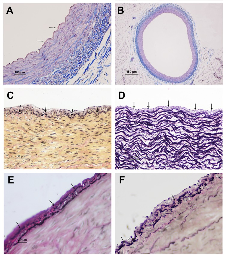
Fig 1. The microscope structures of coronary artery.
A: A monolayer of endothelial cell adhered to internal elastic membrane in 1-day old yak (arrowheads) (Massion trichrome). B: An integral structure of the left circumflex of coronary artery in 1-day old yak (Massion trichrome). C: Internal elastic membrane was disrupted in heart of 6-month old yak (arrowheads) (VVG). D: The internal elastic membrane was divided into two layers in 1-year old yak (arrowheads) (VVG). E: Those internal elastic membrane and smooth muscle cells together form Muscle elastic zone in 2-year old yak (arrowheads) (VVG). F: Secondary internal elastic membrane ruptured, or even fall off, and smooth muscle membrane exposed in old yak (arrowheads) (VVG).

