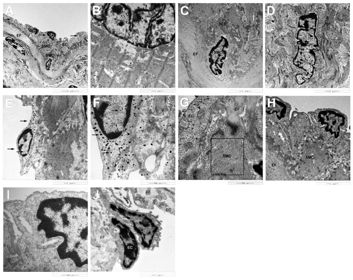
Fig 8. Immunogold labeling of HIF-1α and VEGF protein in the yak heart.
A: Electron microscope images showing HIF-1α immunopositive silver grains in endothelial cells (arrowheads). B: HIF-1α immunopositive silver grains in cardiac muscle cells. C: No HIF-1α immunopositive silver grains in smooth muscle cells. D: Electron microscope images showing VEGF immunopositive silver grains in smooth muscle cells. E: VEGF immunopositive silver grains in endothelial cells (arrowheads). F: High magnification of VEGF immunopositive silver grains in endothelial cells (arrowheads). G: Some fine filaments were found in smooth muscle cells (boxed area). H: Smooth muscle cell has a higher electron density than endothelial cells. I, J: No gold particles were found in control sections. SMC: smooth muscle cell, EC: endothelial cell, EF: elastic fiber, SEF: secondary elastic fiber.

