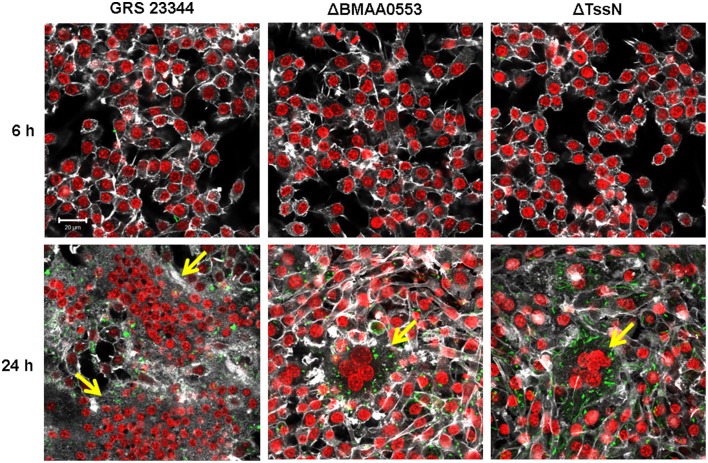
Figure 3.
Multinucleated giant cell (MNGC) formation in macrophage-like cells. RAW 264.7 cells were incubated with GRS 23344 (left), ΔBMAA0553 (middle), or ΔTssN (right) at an MOI of 1 for 6 h (top) and 24 h (bottom) before being stained for actin (white), macrophage nuclei (red) and Bm (green) and then visualized by confocal microscopy. The yellow arrows indicate the presence of a MNGC. Images are representative of three separate experiments. Scale bar = 20 μm.

