
FIG 2.
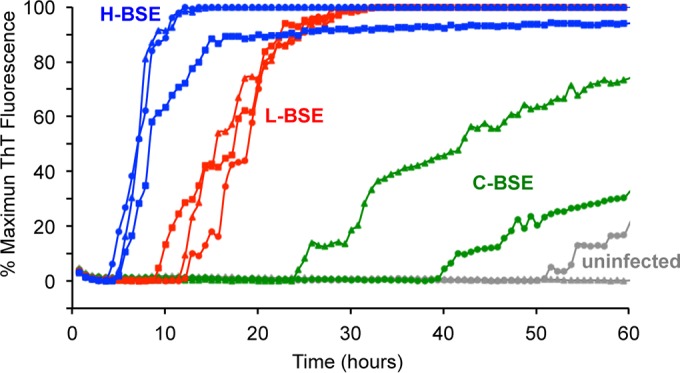
RT-QuIC detection of C-, L-, and H-BSE prion seeding activity using bank vole rPrPSen 23–230, M109. Quadruplicate RT-QuIC reaction mixtures were seeded with 10−5 dilutions of brain tissues from uninfected (gray lines, n = 2), C-BSE-affected (green lines, n = 2), L-BSE-affected (red lines, n = 3) and H-BSE-affected (blue lines, n = 3) cattle. A final SDS concentration of 0.001% in combination with 300 mM NaCl was used with the BV rPrPSen 23–230, M109 substrate. Similar results were observed in three independent experiments, and representative RT-QuIC data are shown. Thioflavin T fluorescence measurements (the average of four replicate wells; y axis) are plotted as a function of time (hours; x axis).