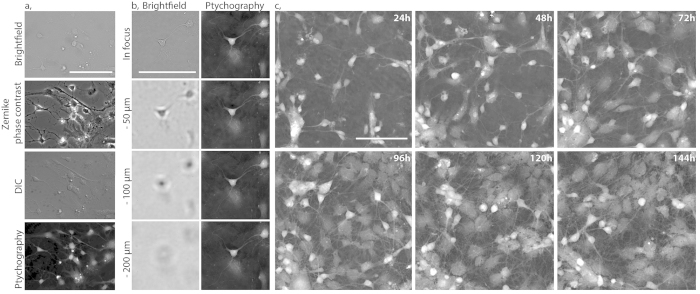
Figure 1. Time-lapse ptychographic phase imaging of primary rat hippocampal neuronal cultures.
(a) Brightfield, Zernike phase contrast, DIC and ptychographic phase image showing the same field of view of primary neuronal cultures. (b) Brightfield images captured on the VL21 microscope acquired at various focal positions by moving objective in the z axis. At each focal position a ptychographic phase image was obtained and successfully refocused post-acquisition. (c) Maturation of primary hippocampal neuron cultures imaged in time-lapse starting at day 1 in-vitro. A 550 × 550 μm field of view was acquired every 6 minutes for a total period of 6 days (144 hours). Representative images show 24 hour cropped snapshots from a continuous time lapse sequence (scale bar = 100 μm, full time lapse sequence available as supplementary video S1).

