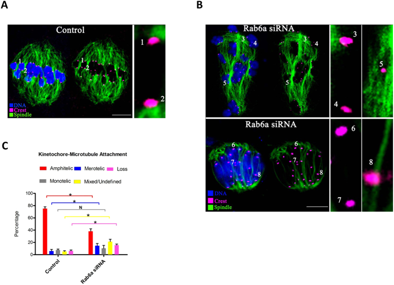
Figure 4. Rab6a-depleted oocytes display impaired kinetochore-microtubule attachments.
(A) Control and Rab6a-siRNA oocytes at MI stage were labeled with α-tubulin antibody to visualize spindle (green), CREST to detect kinetochore (purple), and co-stained with Hoechst 33342 for chromosomes (blue). (A) Representative confocal sections showing the amphitelic attachment in control oocyte (Chromosome 1 and 2). (B) Representative confocal sections showing the monotelic attachment (Chromosome 3 and 4), mixed/undefined attachment (Chromosome 5), loss attachment (Chromosome 6 and 7), and merotelic attachment (Chromosome 8) in Rab6a-siRNA oocytes. (C) Quantitative analysis of K-MT attachments in oocytes as indicated. Kinetochores in regions where fibers were not easily visualized were not included in the analysis. 15 control oocytes and 12 Rab6a-siRNA oocytes were examined respectively. Scale bars, 15 μm. *p < 0.05 vs. controls.

