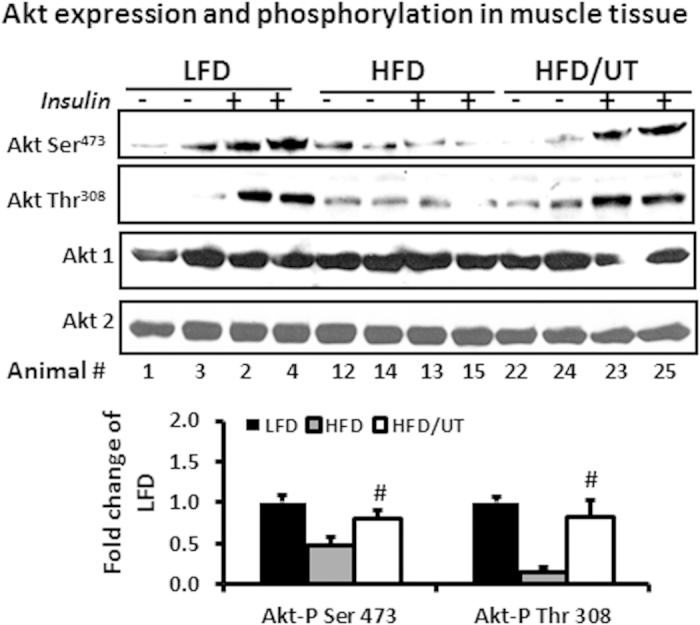
Figure 2. UT extract enhances insulin signaling in skeletal muscle tissue.

Male C57BL/6J mice were fed the experimental diets for 12 weeks. Ten minutes prior to sacrifice, mice were injected with 2 U/Kg body weight insulin or saline. Protein homogenates from the gastrocnemius muscle were separated by SDS-PAGE and analyzed by immune blot analysis. Representative blots for Akt phosphorylation and expression are shown. Fold change relative to LFD alone for each Akt phosphorylation was calculated and mean ± SEM graphed (n = 11). #denotes significant difference between HFD and HFD + UT (P < 0.05).
