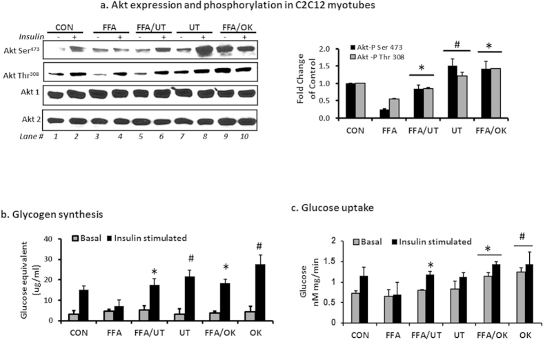
Figure 3. UT extract enhances insulin signaling and glucose uptake in skeletal muscle cells.
Fully differentiated C2C12 myotubes were incubated with palmitate (250 uM) with or without UT (5 ug/ml) or 10 nM okadaic acid (OK) for 16 hours and (a) myotubes were stimulated with vehicle or 100 nM insulin for 8 minutes. Cell lysates were separated by SDS-PAGE and analyzed by immune blot analysis for Akt serine and threonine phosphorylation and Akt protein expression. The bar chart represents quantification of band density by image J software. (b) A second set of cells was used to determine glycogen accumulation with use of glycogen hydrolysis followed by glucose determination in both basal and insulin-stimulated states. Results represent an experiment independently repeated three times on different batches of myotubes. (c) A third set of cells was used to determine glucose uptake in both basal and insulin stimulated states by determining radioactivity after uptake of 2-deoxy glucose. *denotes significant difference with FFA only treatment (P < 0.05). #denotes significant difference with the control treatment (P < 0.05).

