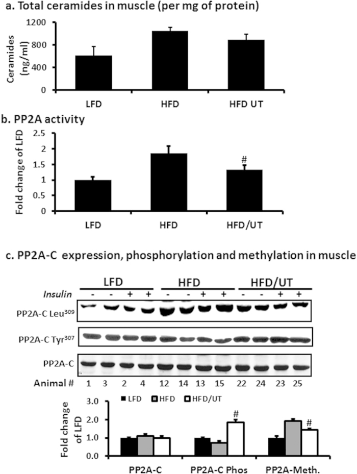
Figure 4. UT extract attenuates HFD induced hyperactivity of PP2A and enhances Akt phosphorylation in skeletal muscle.

Male C57BL/6J mice were fed were fed the experimental diets for 12 weeks. After sacrifice (a) 30 mg of gastrocnemius muscle was extracted for lipids by the Folch partition and ceramide quantities determined by tandemn mass spectrometry (LC/MS/MS). (b) 30 mg muscle was homogenized and lysates used to determine phosphatase PP2A activity by a standard kit (EMD Millipore, Temecula CA). (c) Protein homogenates from 20 mg gastrocnemius muscle were separated by SDS-PAGE and analyzed by immune blot analysis. Representative blots for PP2A- C protein expression, PP2A-C tyrosine phosphorylation and PP2A-C leucine methylation are shown. Fold change relative to LFD alone for each protein was calculated and mean ± SEM graphed (n = 11). #denotes significant difference with HFD (P < 0.05).
