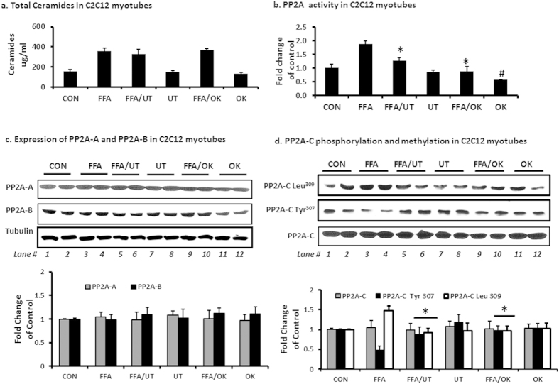
Figure 5. UT extract attenuates FFA induced hyperactivity of PP2A and enhances Akt phosphorylation in C2C12 myotubes.
Fully differentiated C2C12 myotubes were incubated with palmitate (250 uM) with or without UT (5 ug/ml) or 10 nM okadaic acid (OK) for 16 hours. (a) Cells were extracted for lipids by the Folch partition and ceramide quantities determined by tandemn mass spectrometry (LC/MS/MS). (b) Cell lysates were used to determine PP2A activity by a standard kit (EMD Millipore, Temecula CA, USA). (c) Cell lysates were separated by SDS-PAGE and analyzed by immune blot analysis for PP2A-A and PP2A-B protein expression and (d) PP2A-C protein expression, PP2AC tyrosine phosphorylation and PP2A-C leucine methylation. Each panel represents an experiment independently repeated three times on different batches of myotubes. *denotes significant difference with FFA only treatment (P < 0.05). #denotes significant difference with the untreated control treatment (P < 0.05).

